Category: STEM (Science, Technology, Engineering and Mathematics)
ORIGINAL
The utilization of chlorophyll and micro-lead in bio-silicon as a foundation for shielding against X-ray radiation in the medical field
Utilización de clorofila y microplomo en biosilicio como base para el blindaje contra la radiación de rayos X en el ámbito médico
Ahmed Ehsan Jassem1 *, Mohamed Hamzah Al-Maamori2 *, Ahmed Fadhil Hamzah3 *
1Department of Air Conditioning and Refrigeration Techniques Engineering University of Warith Al-Anbiyaa, Karbala, Iraq.
2Departmentof Biomedical Engineering University of Al-Mustaqbal, Iraq.
3Department of Engineering of Polymer and Petrochemical Industries, University of Babylon, Iraq.
Cite as: Jassem AE, Al-Maamori MH, Hamzah AF. The utilization of chlorophyll and micro-lead in bio-silicon as a foundation for shielding against X-ray radiation in the medical field. Salud, Ciencia y Tecnología - Serie de Conferencias. 2024; 3:872. https://doi.org/10.56294/sctconf2024872
Submitted: 06-02-2024 Revised: 15-04-2024 Accepted: 10-06-2024 Published: 11-06-2024
Editor: Dr.
William Castillo-González ![]()
Note: Paper presented at the 3rd Annual International Conference on Information & Sciences (AICIS’23).
ABSTRACT
Shielding aprons were produced using microlead with chlorophyll, which is a viable alternative to the widely used sheet lead aprons for shielding against medical radiation. A study was conducted to examine the effectiveness of five types of radiation shielding sheets composed of a blend of microlead and chlorophyll at varying concentrations (50, 100, 150, 200, and 250 wt% with 35 wt%, respectively) with biosilicon as a base. A comparison was made between the transmission dosages of these sheets and a lead standard (commercial shield). The tensile strength was tested when radiation shielding sheets were being made and used the European Standard for Industry test method (IEC 61331-3:2014) for X-ray protection equipment to measure the transmission dosage. and compare the results with radiation transmitted through a lead standard that had different thicknesses (0,05, 0,1, 0,15, 0,2, 0,25, 0,3, and 0,35 mm). These measurements were taken at tube voltages of 30, 60, 90, 120, 150, and 180 kVp. In the results, it was found that using a mixture of 150 % microlead and 35 % chlorophyll worked to measure a dose similar to 0,3 mm of lead. The transmission dose was 13,58 mR and 13,8 mR for sheet lead, and the density of the shield is 1,72 g/cm3. For this reason, it could be used instead of lead sheets, making it a good choice for protecting medical equipment from radiation.
Keyword: Shield Anti X-Ray; Medical Radiation; Chlorophyll; Micro Lead.
RESUMEN
Se fabricaron delantales de blindaje utilizando microplomo con clorofila, que es una alternativa viable a los delantales de láminas de plomo ampliamente utilizados para el blindaje contra la radiación médica. Se llevó a cabo un estudio para examinar la eficacia de cinco tipos de láminas de blindaje contra la radiación compuestas por una mezcla de microplomo y clorofila en distintas concentraciones (50, 100, 150, 200 y 250 wt% con 35 wt%, respectivamente) con biosilicio como base. Se realizó una comparación entre las dosis de transmisión de estas láminas y un estándar de plomo (blindaje comercial). La resistencia a la tracción se ensayó durante la fabricación de las láminas de blindaje contra la radiación y se utilizó el método de ensayo de la Norma Europea para la Industria (IEC 61331-3:2014) para equipos de protección contra rayos X para medir la dosis de transmisión y comparar los resultados con la radiación transmitida a través de un estándar de plomo que tenía diferentes espesores (0,05, 0,1, 0,15, 0,2, 0,25, 0,3 y 0,35 mm). Estas mediciones se realizaron con tensiones de tubo de 30, 60, 90, 120, 150 y 180 kVp. En los resultados se comprobó que utilizando una mezcla de 150 % de microplomo y 35 % de clorofila se obtenía una dosis similar a la de 0,3 mm
de plomo. La dosis de transmisión fue de 13,58 mR y de 13,8 mR para la lámina de plomo, y la densidad del blindaje es de 1,72 g/cm3. Por este motivo, podría utilizarse en lugar de las láminas de plomo, lo que lo convierte en una buena opción para proteger los equipos médicos de la radiación.
Palabra clave: Escudo Anti Rayos X; Radiación Médica; Clorofila; Microplomo.
INTRODUCTION
The increased advancement of radiation-utilizing medical equipment procedures has led to a growing concern over individual radiation protection and safety. A considerable body of research has been dedicated to investigating the efficacy of medical radiation shielding, primarily driven by the crucial connection between shielding and the potential harm caused by radiation exposure to both patients and professionals involved in radiation-related procedures.(1,2) Aral and colleagues conducted a research investigation to assess the effectiveness of substituting lead with tungsten, bismuth, and barium sulfate granules for X-ray radiation shielding. In observance of medical protection regulations, the X-ray attenuation ratios were determined at tube potentials of 80, 100, and 150 kV. A 1,55 mm-thick coating embedded with bismuth is capable of 90 % attenuation of X-ray radiation at 100 kV, according to the findings of the investigation. An equivalent degree of attenuation is also attained by a tungsten protective coating measuring 1,73 mm in thickness.(3)
In their study, Dong Liang et al. conducted simulations using the Monte Carlo method to evaluate the effectiveness of shields composed of tungsten and bismuth in protecting individuals from X-ray radiation at 150 kV. The shields were tested at a thickness of 2 kg/m2. The findings indicate that the performance of shields made solely from tungsten iron, as a single metal material, surpasses that of shields made from bismuth material. In the event of a double-layer metal of equivalent quality, it can be argued that tungsten-bismuth exhibits superior characteristics compared to bismuth-tungsten. The protective efficacy is enhanced when the ratio of metal-mixed tungsten to bismuth is 0,5:0,5 or when the tungsten ratio is relatively high.(4)
Seon-Chil Kim and colleagues have fabricated the use of barium sulfate as a base material for shielding against tungsten, molybdenum, rubber, and silicon at different tube voltages ranging from 50 to 150 kVp. Based on the findings, it was observed that the amalgamation of barium, tungsten, molybdenum, and silicon yields an equivalent proportion of lead at a measurement of 0,3 mm. Hence, it possesses the potential to serve as a substitute for lead sheets, thereby qualifying as a suitable sheet for medical radiation shielding.(5,6)
In their study, Shruti Nambiar et al. undertake an investigation and put forth a proposal for the fabrication of shields comprised of polydimethylsiloxane (PDMS) with varying weight percentages of micropowderized bismuth oxide (BO). The results of the experiment demonstrated that the PDMS/BO nanocomposite shield, comprising 44,44 weight percent BO and measuring 3,73 mm in thickness, successfully attenuated the intensity of every scattered X-ray generated at a tube potential of 60 kV.(7,8)
In a study conducted by Nurul Z. et al., composite materials for X-ray shielding were fabricated utilizing PbO, epoxy, and Pb3O4. Higher density was positively correlated with the composites' capacity to reduce attenuation, according to the findings. Furthermore, it was noted that the optimal dispersion of fillers within the epoxy matrix took place when the mean particle size ranged from 1 to 5 microns for composites containing filler loadings not exceeding 30 weight percent and from 5 to 15 microns for composites containing filler loadings not exceeding 50 weight percent.(9,10)
The study conducted by Botelho et al. centered on the analysis of X-ray transmission characteristics across materials measuring between the microscale and nanoscale. A concentration of 5 % was applied to beeswax containing CuO nanoparticles (mean grain size: 13,4 nm) and CuO microparticles (mean particle size: 56 mm) individually in this experiment. The results of the research indicate that X-ray beams generated at tube voltages of 60 and 102 kV continue to transmit without interruption when traversing plates made of the specified materials, which include CuO at the micro- and nanoscales. The X-rays are significantly attenuated by the nanostructured CuO plates, which effectively reduce the intensity by a minimum of 14 % for radiation beams produced at tube voltages of 26 and 30 kV. The discrepancy observed in the low energy range can be attributed to two elements: the increased particle density per gram in the plates engineered with CuO nanoparticles and the influence of grain size on the transmission of X-rays.(11,12)
Seon-Chil Kim conducted research on developing protective clothing made from polyethylene terephthalate (PET) fiber. The garment was constructed utilizing environmentally friendly components, specifically BaSO4 and Bi2O3. Yarns were produced through the process of melt spinning, and garments were created by weaving. containing 5 weight percent of shielding material. The average radiation shielding of the fibers was 9–13 %, with the Bi2O3 fiber exhibiting superior shielding efficacy compared to the BaSO4 fiber. The strands created from this material are utilized to create a shield that offers protection against low-dose and dispersed rays in medical applications, as well as for aerospace radiation protection.(13)
Seon-Chil Kim investigated a shield product composed of a blend of liquid polymer and tungsten, as well as a fillet created by combining the same material and then laminating it using a heat-treatment press following the injection process. The tungsten content and thickness were identical, with a weight percentage of 85 % and a measurement of 0,3 mm, respectively. The abalone shell-based shielding film, with its laminated structure, exhibited a shielding rate in the high-energy zone of over 7 %. The shielding film demonstrated a 16 % lower shielding ratio at 120 kVp compared to a 0,3 mm lead plate, thus verifying the radiation-shielding effectiveness of the layered-structure shielding film.(14)
The present work employed microlead containing chlorophyll as an alternative to conventional lead sheets in order to fabricate a radiation shielding sheet that is more environmentally sustainable. Additionally, various components were incorporated into the microlead-chlorophyll mixture to enhance its limited miscibility. The objectives of this study were to devise a composition and methodology aimed at improving the rate of shielding as well as assess its efficacy. Furthermore, the optimal mixing composition was ascertained by quantifying the radiation doses at 30-180 kVp in order to evaluate the shielding efficacy of recently produced sheets made from lead.
Materials Used
In this proposed shield, we will use silicone rubber MM928 (RTV 754) from CHT Germany GmbH, which is considered a binder or base material, as well as micro-lead powder 2,5 µm and chlorophyll extracted from chlorophyll-rich plants such as celery.
Preparation Samples
Five distinct varieties of radiation shielding sheets were produced through the combination of PEG 400, chlorophyll, elastomer silicon, and micro-lead. Where chlorophyll is extracted from plants rich in it, such as celery and others, by means of a xylene solvent, the celery is soaked in xylene for twenty-four hours, after which the impurities are filtered, and then the extract is dried to obtain chlorophyll powder.
Then the amount of chlorophyll is weighed to obtain a percentage of 35 %, and then microscopic lead is added to the chlorophyll in different proportions (50, 100, 150, 200, 250) wt%, and the micro lead is mixed with chlorophyll using a small amount of xylene using ultrasound waves for 20 minutes. The silicone rubber is modified by adding 5 % wt% of PEG 400 to increase hydrophilicity.(15) The suspension is added to the modified silicone and mixed with a magnetic mixer for six hours, after which the hardener is added to the silicone and mixed again with the magnetic mixer for three minutes. The mixture is cast into a glass mold with dimensions of 15 x 15 cm, and a vacuum is made for a quarter of an hour to remove bubbles in the shield, then placed in the oven for an hour at a temperature of 50 Co and a pressure of 5 bar, after which it is extracted from the oven and left to harden to obtain a silicone shield. Figure 1 illustrates a radiation shielding sheet composed of silicon, which serves as a finished product. The dimensions of the sample are 150 mm x 150 mm x 2 mm.
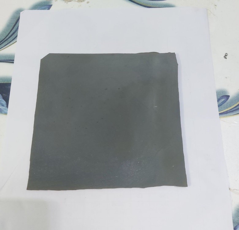
Figure 1. Radiation shielding sheet composed of silicon
To determine whether the shielding sheet is effective, a lead equivalent test method is utilized,(17,18) specified in the European Industrial Standard for X-ray protective supplies was employed (IEC 61331-3:2014).(19) Additionally, the GE Healthcare DefinirionTM 6000 X-ray generator's effective energy was measured (USA). The geometric parameters essential for the evaluation of effective energy are illustrated in figure 2. In this investigation, the tube current was set at 200 mA, and the exposure period was maintained at 0,1 s. The collimator featured a fixed filtration of 1,5 mmAl with an additional filtration of 1,5 mmAl, whereas the X-ray tube featured intrinsic filtration settings of 0,1 mmBe and 4,0 mmBe. In accordance with the clinically accepted range, the tube voltage was modified to a variety of kilovolt peak (kVp) values: 30, 60, 90, 120, 150, and 180 kVp. By modifying the aluminum absorber's breadth, the half-value layer could be ascertained. A calibrated and tested ion chamber, more specifically the QUART didoNEO R model, was utilized to determine the exposure dose. At present, when employing an ionization dosimeter to accurately determine the exposure dose at a pressure of 1 atm and a temperature of 22 Co.
The linear absorption coefficient (µ value) was acquired through the computation of the slope of the y-axis graph, which was utilized to ascertain the half-value layer. By applying the logarithm to the exponential equation for attenuation (I = I0e-µx),(20) The formula utilized to determine the half-value layer is given by half-value layer = 0,693/µ. Hubbell's mass absorption coefficient table was employed to compute the effective energy of the half-value layer associated with a specific energy,(21) in order to determine the effective energy for efficient energy utilization.(5,22) Figure 3 illustrates the geometric parameters that were considered during the performance testing of the shielding sheets that were produced. By conducting a direct comparison between the transmission doses and lead equivalents, the capability effectively showcased the effectiveness of the shielding. The radiation exposures were quantified by placing a shield between the ionization dosimeter and the X-ray beam. The experiment was conducted 10 different times, and the mean value was utilized for the purpose of analysis. In order to compare the shielding ability, experiments were conducted to examine the transmission dosages of lead standards measuring 0,05, 0,1, 0,15, 0,2, 0,25, 0,3, and 0,35 mm.(23)

Figure 2. A diagram is employed to measure the half-value layer, which provides information on the effective energy (5,24)
RESULT AND DISCUSSION
The shielding sheets fabricated in this study possess a thickness of 2 mm. Table 1 presents the recorded values for tensile strength (MPa) and density (g/cm3). The chlorophyll does not greatly affect the mechanical properties of the manufactured shield, but it does slightly reduce the mechanical properties such as tensile strength, and the increase in tensile and density properties is caused by the microscopic lead added to the panels. Table 2 presents the pertinent characteristics about the quality and efficacy of the X-ray used for assessing the shielding rate. The shielding rate was evaluated by using microlead with chlorophyll properties at a tube voltage of 60 kilovolts peak (kVp). The linear absorption coefficient and half-value layer, without further filtration, were determined to be 0,3134 per millimeter and 2,40 mmAl, respectively. Therefore, the precise value for the effective energy was calculated to be 30,58 kiloelectron volts (keV) by using the mass absorption coefficient table provided by Hubbell.(22,26) Consequently, the effective energy may be enhanced by increasing the tube voltage and including thicker filtering, as these factors influence the quality of X-rays. The shielding capacity of the sheets was evaluated based on the resultant X-ray quality.

Figure 3. An instructional diagram illustrates a measuring configuration intended to assess the efficacy of a radiation shielding sheet. The present arrangement conforms to the lead equivalent testing methodology that is defined in the European Industrial Standard for X-ray protective equipment(17,25)
|
Table 2. Properties of the sheet products |
||
|
Sheet |
Density [g/cm3] |
Tensile strength [MPa] |
|
Non |
1,45 |
85 |
|
B |
1,49 |
140 |
|
C |
1,64 |
200 |
|
D |
1,72 |
270 |
|
E |
1,9 |
356 |
|
F |
2,1 |
320 |
|
Table 3. Comparison between the effective energy and the voltage of the tube. |
||||
|
Tube voltage [kVp] |
Inherent filter |
Added filter |
Half value [mmAl] |
Effective energy [keV] |
|
30 |
1,0 mmBe |
|
0,36 |
19,9 |
|
60 |
4,0 mmBe + 1,5 mmAl |
|
2,4 |
30,6 |
|
90 |
|
4,07 mmAl +0,31 mmCu |
4,91 |
48,3 |
|
120 |
|
4,07 mmAl +0,31 mmCu |
4,419 |
66 |
|
150 |
|
4,07 mmAl +0,31 mmCu |
10,26 |
76,7 |
|
180 |
|
4,07 mmAl +0,31 mmCu |
12,312 |
92,03 |
The comparative investigation included assessing the efficacy of both the traditional lead standard and the recently produced shielding sheets in terms of their capacity to provide protection. The lead standard's shielding capability was determined using the same methodology as previously documented. Table 4 displays the transmission dosages of conventional lead. The research found a direct association between the shielding capacity and the thickness of the lead, indicating that greater thickness or bigger quantities of lead demonstrated stronger shielding capabilities. This study presents the mean transmission doses and lead equivalent values of the shielding sheets produced, aiming to elucidate their effectiveness in shielding in comparison to conventional lead.
|
Table 4. The standard lead transmission doses. |
||||||||
|
Dose (kVp) |
mmPb |
|||||||
|
|
NON |
0,05 |
0,1 |
0,15 |
0,2 |
0,25 |
0,3 |
0,35 |
|
30 |
6,3612 |
1,266 |
0,424 |
0,148 |
0,08 |
0,031 |
0,013 |
0,004 |
|
60 |
18,735 |
5,574 |
2,802 |
1,465 |
1,119 |
0,668 |
0,481 |
0,305 |
|
90 |
44,435 |
18,4 |
11,195 |
6,7 |
5,61 |
3,7 |
2,8 |
1,915 |
|
120 |
75,9 |
36,17 |
25,46 |
16,3 |
14,1 |
9,6 |
7,5 |
5,3 |
|
150 |
112,21 |
59,01 |
41,23 |
28,14 |
25,15 |
17,5 |
13,8 |
9,8 |
|
180 |
134,652 |
70,812 |
49,476 |
33,768 |
30,180 |
21,000 |
16,560 |
11,760 |
|
Note: The unit of measurement is mR. |
||||||||
The shielding efficacy was enhanced by the incorporation of lead microparticles as an alternative to sheet lead. This was achieved by combining lead microparticles with 35 % chlorophyll and 5 % polyethylene glycol in silicone rubber, therefore reducing the inter-particle porosity. In general, chlorophyll absorbs light within the range of 400–700 nm and converts it into energy, but when it shines high energy, such as X-rays, with a short wavelength, chlorophyll absorbs it and dissipates it to heat due to the carotenoids in chloroplasts, so the radiation penetrating from the panels manufactured from them decreases,(27) in addition to the presence of microscopic lead that disperses energy that chlorophyll could not dissipate. Furthermore, the process of vacuum defoaming was employed in a sequential manner prior to the hardening stage in order to enhance the packing of particles and improve the miscibility inside the silicon resin. The transmission dosages are presented in Table 5, which classifies them according to the thickness of the lead standard and the composition of the shielding sheets utilized. The sheet with a composition of 150 % lead and 35 % chlorophyll exhibited identical shielding effectiveness to a 0,3 mm lead equivalent at 150 kVp. Furthermore, the sheet composed of 200 % lead and 35 % chlorophyll likewise showed comparable shielding ability to a 0,3 mm lead equivalent at 150 kVp.
|
Table 5. A comparison between radiation shielding sheeting and transmission doses Dose (kVp) |
|||||||
|
|
Non |
B |
C |
D |
E |
F |
|
|
30 |
Mean |
6,361 |
0,783 |
0,573 |
0,189 |
0,122 |
0,034 |
|
|
mmPb |
0,000 |
0,039 |
0,063 |
0,095 |
0,134 |
0,236 |
|
60 |
Mean |
18,735 |
2,189 |
1,040 |
0,368 |
0,225 |
0,076 |
|
|
mmPb |
0,000 |
0,036 |
0,059 |
0,092 |
0,121 |
0,221 |
|
90 |
Mean |
44,435 |
6,083 |
4,897 |
3,841 |
2,797 |
2,183 |
|
|
mmPb |
0,000 |
0,033 |
0,056 |
0,089 |
0,120 |
0,219 |
|
120 |
Mean |
75,9 |
10,179 |
9,747 |
8,641 |
7,981 |
7,754 |
|
|
mmPb |
0,000 |
0,033 |
0,052 |
0,084 |
0,117 |
0,215 |
|
150 |
Mean |
112,21 |
15,750 |
14,982 |
13,58 |
12,58 |
11,981 |
|
|
mmPb |
0,000 |
0,032 |
0,051 |
0,086 |
0,119 |
0,217 |
|
180 |
|
134,652 |
18,940 |
17,984 |
16,897 |
15,798 |
15,012 |
|
|
mmPb |
0,000 |
0,032 |
0,053 |
0,086 |
0,118 |
0,218 |
|
Note: The unit of measurement is mR. |
|||||||
The use of radiation in medical technology and equipment has significantly contributed to the diagnosis and treatment of several ailments. The application of radiation in invasive medical procedures, such as angiography, has witnessed significant growth, resulting in an escalation of radiation exposure for both patients and healthcare personnel. Consequently, ensuring individual radiation protection has emerged as a crucial concern.
In contemporary medical discourse, the primary constituent of shielding materials is lead. Consequently, aprons crafted from micro-lead and chlorophyll have gained prominence as a cost-effective, easily manufacturable, and pleasant alternative. This is attributed to the inherent flexibility of the sheet, which not only enhances comfort but also mitigates toxicity concerns. Furthermore, the process of mass manufacture and dissemination poses significant challenges.
The lead equivalence of current lead aprons ranges from 0,25 to 0,5 mm. A lead apron with a thickness of 0,5 mmPb has a shielding effectiveness of over 95 % against direct radiation at 100 kVp. Hence, it is imperative to have a minimum shielding thickness of 0,3 mmPb in order to account for the reduction in shielding efficacy. Previous studies have demonstrated that micro-lead, when combined with chlorophyll at concentrations of 150 % and 35 %, respectively, exhibits a remarkable capacity to protect against various forms of medical radiation. This finding is visually represented in figure 4. One of the challenges associated with this particular shielding method is the intricate dispersion of micro-lead, a process that necessitates a significant amount of time for adequate preparation prior to commencing mass production. Furthermore, achieving optimal shielding and efficiency necessitates a reduction in lead particle size.
The present investigation resulted in the development of a protective barrier against x-ray radiation. Within the realm of medicine, a comparative analysis is conducted between bio-silicon, possessing a thickness of 2 mm and exhibiting a relatively low weight, and the existing lead sheet shields with a thickness of 0,3 mm that are presently employed in healthcare facilities. The researchers considered the effectiveness of the shield in environments with high levels of radiation and determined that a plate thickness of 1 cm is sufficient to provide protection against low radiation fields.
Figure 4 show a diagram demonstrates a comparison of the radiation shielding sheet, focusing on the shielding capacity of microlead, chlorophyll, and lead in the 40–80 keV X-ray energy region for diagnostic purposes. It was discovered that the absorption of microlead with chlorophyll was higher in comparison to that of standard lead.

Figure 4. Diagram demonstrates a comparison of the radiation shielding sheet
CONCLUSION
A medical radiation shielding sheet was produced by incorporating varying concentrations (50, 100, 150, 200, and 250 wt%) of microlead and chlorophyll (at a constant concentration of 35 wt%) into biosilicon. This approach proved to be more cost-effective compared to previously suggested shielding methods, while still achieving a comparable shielding effect to that of lead. The radiation attenuation properties of shielding sheets with a thickness of 2 mm were evaluated at tube voltages of 30, 60, 90, 120, 150, and 180 kilovolts peak (kVp). The use of a composite consisting of microlead at a concentration of 150 wt% in conjunction with chlorophyll at a concentration of 35 wt% showed notable efficacy in accurately measuring a dosage at 150 Kvp of 13,58 mR and 3,84 mR at 90 Kvp, which also exhibited a remarkably high tensile strength. The shielding efficacy of the micro-lead composite, consisting of 150 wt% lead and 35 wt% chlorophyll, was shown to be comparable to that of a 0,3 mm lead equivalent.
REFERENCES
1. S. G. Barnard, E. A. Ainsbury, R. A. Quinlan, and S. D. Bouffler, “Radiation protection of the eye lens in medical workers—basis and impact of the ICRP recommendations,” The British Journal of Radiology, vol. 89, no. 1060, p. 20151034, Apr. 2016, doi: 10.1259/bjr.20151034.
2. R. Najjar, “Radiology’s Ionising Radiation Paradox: Weighing the Indispensable Against the Detrimental in Medical Imaging,” Cureus, Jul. 2023, doi: 10.7759/cureus.41623.
3. N. Aral, F. Banu Nergis, and C. Candan, “An alternative X-ray shielding material based on coated textiles,” Textile Research Journal, vol. 86, no. 8, pp. 803–811, May 2016, doi: 10.1177/0040517515590409.
4. D. Liang, F. Shen, Z. Bao, Y. Liu, and H. Li, “Research on textile materials for X-ray shielding,” E3S Web of Conferences, vol. 290, p. 01013, Jul. 2021, doi: 10.1051/e3sconf/202129001013.
5. S.-C. Kim, K.-R. Dong, and W.-K. Chung, “Medical radiation shielding effect by composition of barium compounds,” Annals of Nuclear Energy, vol. 47, pp. 1–5, Sep. 2012, doi: 10.1016/j.anucene.2012.04.014.
6. L. Yu, P. L. Yap, A. Santos, D. Tran, and D. Losic, “Lightweight polyester fabric with elastomeric bismuth titanate composite for high-performing lead-free X-ray shielding,” Radiation Physics and Chemistry, vol. 205, p. 110726, 2022, doi: 10.1016/j.radphyschem.2022.110726.
7. S. Nambiar, E. K. Osei, and J. T. W. Yeow, “Polymer nanocomposite-based shielding against diagnostic X-rays,” Journal of Applied Polymer Science, vol. 127, no. 6, pp. 4939–4946, Mar. 2013, doi: 10.1002/app.37980.
8. A. Trajkovska Petkoska, “Assessment of the Attenuation Properties of Commercial Lead-Free Radiation-Shielding Composite Materials Against Medical X-rays,” Journal of Composites Science, vol. 7, no. 10, p. 424, Oct. 2023, doi: 10.3390/jcs7100424.
9. N. Z. N. Azman, S. A. Siddiqui, R. Hart, and I. M. Low, “Microstructural design of lead oxide-epoxy composites for radiation shielding purposes,” Journal of Applied Polymer Science, vol. 128, no. 5, pp. 3213–3219, Jun. 2013, doi: 10.1002/app.38515.
10. M. J. R. Aldhuhaibat, M. S. Amana, N. J. Jubier, and A. A. Salim, “Improved gamma radiation shielding traits of epoxy composites: Evaluation of mass attenuation coefficient, effective atomic and electron number,” Radiation Physics and Chemistry, vol. 179, p. 109183, Feb. 2021, doi: 10.1016/j.radphyschem.2020.109183.
11. M. Z. Botelho, R. Künzel, E. Okuno, R. S. Levenhagen, T. Basegio, and C. P. Bergmann, “X-ray transmission through nanostructured and microstructured CuO materials,” Applied Radiation and Isotopes, vol. 69, no. 2, pp. 527–530, Feb. 2011, doi: 10.1016/j.apradiso.2010.11.002.
12. B. Bazi et al., “Trace-element analysis of mineral grains in Ryugu rock fragment sections by synchrotron-based confocal X-ray fluorescence,” Earth, Planets and Space, vol. 74, no. 1, p. 161, Nov. 2022, doi: 10.1186/s40623-022-01726-y.
13. S.-C. Kim, “Construction of a Medical Radiation-Shielding Environment by Analyzing the Weaving Characteristics and Shielding Performance of Shielding Fibers Using X-ray-Impermeable Materials,” Applied Sciences, vol. 11, no. 4, p. 1705, Feb. 2021, doi: 10.3390/app11041705.
14. S.-C. Kim, “Medical-Radiation-Shielding Film Fabricated by Imitating the Layered Structure Pattern of Abalone Shell and Verification of Its Shielding Effect,” Materials, vol. 16, no. 24, p. 7700, Dec. 2023, doi: 10.3390/ma16247700.
15. T. Vladkova, “Surface modification of silicone rubber with poly(ethylene glycol) hydrogel coatings,” Journal of Applied Polymer Science, vol. 92, no. 3, pp. 1486–1492, May 2004, doi: 10.1002/app.20001.
16. K. A. Montoya-Villegas et al., “Controlled surface modification of silicone rubber by gamma-irradiation followed by RAFT grafting polymerization,” European Polymer Journal, vol. 134, p. 109817, Jul. 2020, doi: 10.1016/j.eurpolymj.2020.109817.
17. T. Schöpf and T. Pichler, “Radiation Protection Clothing in X-Ray Diagnostics – Influence of the Different Methods of Measurement on the Lead Equivalent and the Required Mass,” RöFo - Fortschritte auf dem Gebiet der Röntgenstrahlen und der bildgebenden Verfahren, vol. 188, no. 08, pp. 768–775, Jun. 2016, doi: 10.1055/s-0042-106651.
18. R. S. Livingstone and A. Varghese, “A simple quality control tool for assessing integrity of lead equivalent aprons,” Indian Journal of Radiology and Imaging, vol. 28, no. 02, pp. 258–262, Apr. 2018, doi: 10.4103/ijri.IJRI_374_17.
19. T. B. S. Institution, BSI Standards Publication Protective devices against diagnostic medical, Three. BSI Standards Limited, 2014.
20. F. H. Omoumi, X. Wu, M. U. Ghani, M. D. Wong, Y. Li, and H. Liu, “Mathematical estimation of half‐value layer thicknesses,” Journal of Applied Clinical Medical Physics, vol. 22, no. 10, pp. 320–328, Oct. 2021, doi: 10.1002/acm2.13385.
21. T. Kaur, J. Sharma, and T. Singh, “Experimental evaluation of gamma rays shielding parameters for Zn-Cd-Sn-Pb quaternary alloy,” Radiation Physics and Chemistry, vol. 156, pp. 193–198, Mar. 2019, doi: 10.1016/j.radphyschem.2018.11.010.
22. J. H. Hubbell, “Photon mass attenuation and energy-absorption coefficients,” The International Journal of Applied Radiation and Isotopes, vol. 33, no. 11, pp. 1269–1290, Nov. 1982, doi: 10.1016/0020-708X(82)90248-4.
23. H. Çetin, A. Yurt, and S. H. Yüksel, “THE ABSORPTION PROPERTIES OF LEAD-FREE GARMENTS FOR USE IN RADIATION PROTECTION,” Radiation Protection Dosimetry, p. ncw004, Feb. 2016, doi: 10.1093/rpd/ncw004.
24. A. H. Alsaab and S. Zeghib, “Analysis of X-ray and gamma ray shielding performance of prepared polymer micro-composites,” Journal of Radiation Research and Applied Sciences, vol. 16, no. 4, p. 100708, Dec. 2023, doi: 10.1016/j.jrras.2023.100708.
25. R. R. Wargo, A. F. Aljabal, and P. P. Lin, “Evaluation and verification of a simplified lead equivalency measurement method,” Journal of Applied Clinical Medical Physics, vol. 21, no. 2, pp. 152–156, Feb. 2020, doi: 10.1002/acm2.12810.
26. B. Akça and S. Z. Erzeneoğlu, “The Mass Attenuation Coefficients, Electronic, Atomic, and Molecular Cross Sections, Effective Atomic Numbers, and Electron Densities for Compounds of Some Biomedically Important Elements at 59.5 keV,” Science and Technology of Nuclear Installations, vol. 2014, pp. 1–8, 2014, doi: 10.1155/2014/901465.
27. A. Kume, “Importance of the green color, absorption gradient, and spectral absorption of chloroplasts for the radiative energy balance of leaves,” Journal of Plant Research, vol. 130, no. 3, pp. 501–514, May 2017, doi: 10.1007/s10265-017-0910-z.
FINANCING
None.
CONFLICT OF INTEREST
None.
AUTHORSHIP CONTRIBUTION
Conceptualization: Ahmed Ehsan Jassem, Mohamed Hamzah Al-Maamori, Ahmed Fadhil Hamzah.
Research: Ahmed Ehsan Jassem, Mohamed Hamzah Al-Maamori, Ahmed Fadhil Hamzah.
Writing - original draft: Ahmed Ehsan Jassem, Mohamed Hamzah Al-Maamori, Ahmed Fadhil Hamzah.
Writing - revision and editing: Ahmed Ehsan Jassem, Mohamed Hamzah Al-Maamori, Ahmed Fadhil Hamzah.